Nucleus In Plant Cell Under Microscope : Cell Nucleus Function Structure And Under A Microscope Rs Science - This is the best microscope to use because the microscope shows a significant amount of detail the structure that is seen is the cell wall.
byGertrud Landt-0
Nucleus In Plant Cell Under Microscope : Cell Nucleus Function Structure And Under A Microscope Rs Science - This is the best microscope to use because the microscope shows a significant amount of detail the structure that is seen is the cell wall.. It is sometimes hard to see. Describe the nucleus of a cell. They got the name chromatin (gr. Through the microscope, the nucleolus looks like a large dark spot within the nucleus. Look for cells where the nucleus has a consistent color overall.
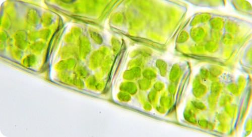
The granulated area is the cell cytoplasm while the huge round part is the nucleus. It is thought that red blood cells evolved without a nucleus in order to maximize space for carrying hemoglobin. Additionally, certain blood disorders can lead to abnormalities in the nuclei, meaning that analysis of the shape and structure of nuclei in blood cells. Search for plant cell nucleus in these categories. (7) parts of nucleus visible only under electron microscopes (don't get me wrong, the nucleus is still visible under light microscopes, just that individual parts of it are only visible under electron microscope) all of the organelles in plant and animal cells can be seen under a light microscope.
The Unit Of Life from www1.biologie.uni-hamburg.de Like animal cell nuclei, this cell nucleus will retain a spherical shape if there is enough room. It is thought that red blood cells evolved without a nucleus in order to maximize space for carrying hemoglobin. Plant cells need protection against variations in temperature, high wind speed, atmospheric moisture etc. Want to learn more about it? You can specify conditions of storing and accessing cookies in your browser. A nucleus can be easily visualized under a compound microscope. The cell nucleus contains all of the cell's genome, except for the small amount of mitochondrial dna and, in plant cells, plastid dna. Plant cell organelles that are invisible under a compound light microscope include mitochondria, ribosomes, endoplasmic reticula.
Image:plant cell seen under electron microscope.
This section on microscopy is meant as an introduction as learners will need to be able to use microscopes later in this cells are microscopic and can only be seen under a microscope. The granulated area is the cell cytoplasm while the huge round part is the nucleus. When viewed under the microscope, the nucleus will appear as a spherical, blue structure surrounded by cytokeratin intermediate filament network. Cell — structure and functions. Centrioles are structures made of microtubules that help organize the mitotic spindle, and in plant cells, when cytokinesis, or the moment two daughter cells form from a single cell, occurs, a cell wall forms between the two cells. Plant cells do not have centrioles like animal cells, just centrosomes. A nucleus can be easily visualized under a compound microscope. The innermost fibrillar centers (fcs), surrounded. Nucleolus is responsible for manufacture of ribosomes while chromatin contains hereditary materials. Oftentimes in plant cells, the central 3. (7) parts of nucleus visible only under electron microscopes (don't get me wrong, the nucleus is still visible under light microscopes, just that individual parts of it are only visible under electron microscope) all of the organelles in plant and animal cells can be seen under a light microscope. A nucleus may contain up to four nucleoli, but within each species. A cell is a very tiny structure which exists in living bodies.
It is also known as nuclear reticulum. It also has a very high resolving power. Additionally, certain blood disorders can lead to abnormalities in the nuclei, meaning that analysis of the shape and structure of nuclei in blood cells. Like animal cell nuclei, this cell nucleus will retain a spherical shape if there is enough room. The innermost fibrillar centers (fcs), surrounded.
Animal Vs Plant Cells Similarities Differences Chart And Examples Rs Science from rsscience.com The granulated area is the cell cytoplasm while the huge round part is the nucleus. Microscopy is a fun and fascinating hobby and i hope that i can share my enthusiasm with you. When observed under the electron microscope, the nucleolus can be seen to consist of three distinguishable regions: The genetic material is dispersed in the cytoplasm without any nuclear membrane. Like animal cell nuclei, this cell nucleus will retain a spherical shape if there is enough room. The nucleus in a photograph of a cell measures 3 mm across. This section on microscopy is meant as an introduction as learners will need to be able to use microscopes later in this cells are microscopic and can only be seen under a microscope. Chroma=color) due to their colorful nature during cell staining when it is viewed under microscope.
When observed under the electron microscope, the nucleolus can be seen to consist of three distinguishable regions:
The cell nucleus contains all of the cell's genome, except for the small amount of mitochondrial dna and, in plant cells, plastid dna. When looking at stained nuclei under a microscope, you notice that some appear uniformly colored, while other appear almost empty, with most of the color. A nucleus may contain up to four nucleoli, but within each species. Chroma=color) due to their colorful nature during cell staining when it is viewed under microscope. They are all typical elements of a cell. Microscopy is a fun and fascinating hobby and i hope that i can share my enthusiasm with you. Microscopes are used to study cells. It also has a very high resolving power. The nucleus was the first organelle to be discovered. The cell outline and the nucleus with its internal structure are clearly visible, as are a series of dark, spherical particles in the cytoplasm. Additionally, certain blood disorders can lead to abnormalities in the nuclei, meaning that analysis of the shape and structure of nuclei in blood cells. Cell — structure and functions. When observed under the electron microscope, the nucleolus can be seen to consist of three distinguishable regions:
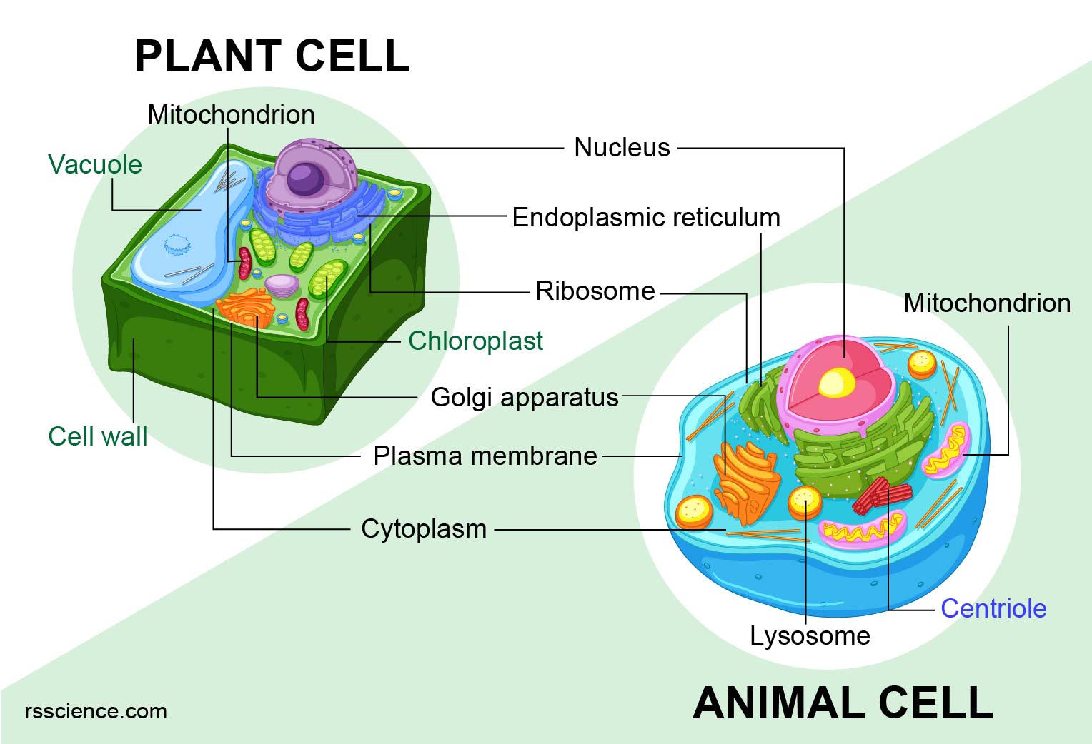
Plant cell organelles that are invisible under a compound light microscope include mitochondria, ribosomes, endoplasmic reticula. As a plant cell what extra layer must be formed to in the onion to separate the daughter cells? Area containing cytosol (mixture of water, salts and organic molecules). In most plant cells, the organelles that are visible under a compound light microscope are the cell wall, cell membrane, cytoplasm, central vacuole, and nucleus. He observed a lumen, the nucleus, in the red blood cells of salmon.
Plant Cell Structure Read Biology Ck 12 Foundation from dr282zn36sxxg.cloudfront.net As a plant cell what extra layer must be formed to in the onion to separate the daughter cells? This section on microscopy is meant as an introduction as learners will need to be able to use microscopes later in this cells are microscopic and can only be seen under a microscope. Observing cells under a microscope. A nucleus may contain up to four nucleoli, but within each species. Search for plant cell nucleus in these categories. This keeps the shape of the cell and is only found in plant under a x400 light microscope we could see the cell wall, cell membrane, nucleus and cytoplasm The cell nucleus contains all of the cell's genome, except for the small amount of mitochondrial dna and, in plant cells, plastid dna. When looking at stained nuclei under a microscope, you notice that some appear uniformly colored, while other appear almost empty, with most of the color.
A cell is a very tiny structure which exists in living bodies.
When observed under the electron microscope, the nucleolus can be seen to consist of three distinguishable regions: Microscope slide cover slip onion. Microscopy is a fun and fascinating hobby and i hope that i can share my enthusiasm with you. If the cell is allowed to yield under pressure and doesn't have to keep its shape completely, the. Inside the nucleus is the nucleolus, which is a dense spherical region in the nucleus. As a plant cell what extra layer must be formed to in the onion to separate the daughter cells? In most plant cells, the organelles that are visible under a compound light microscope are the cell wall, cell membrane, cytoplasm, central vacuole, and nucleus. Interpretation of electron micrographs to identify organelles and deduce the function of specialised cells. Chroma=color) due to their colorful nature during cell staining when it is viewed under microscope. He observed a lumen, the nucleus, in the red blood cells of salmon. (7) parts of nucleus visible only under electron microscopes (don't get me wrong, the nucleus is still visible under light microscopes, just that individual parts of it are only visible under electron microscope) all of the organelles in plant and animal cells can be seen under a light microscope. Want to learn more about it? When viewed under the microscope, the nucleus will appear as a spherical, blue structure surrounded by cytokeratin intermediate filament network.
This is the best microscope to use because the microscope shows a significant amount of detail the structure that is seen is the cell wall nucleus in plant cell. Microscope slide cover slip onion.


Post a Comment