Plant Cell With Microscope - Animal And Plant Cells Microscope Slide Set Microscope Sample Slides Amazon Com Industrial Scientific - Structure of animal cell and plant cell under microscope + diagrams.
byGertrud Landt-0
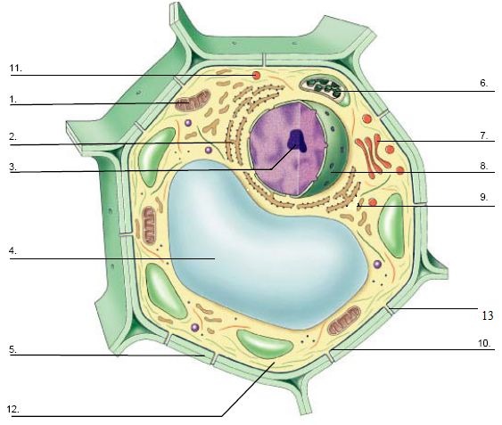
Plant Cell With Microscope - Animal And Plant Cells Microscope Slide Set Microscope Sample Slides Amazon Com Industrial Scientific - Structure of animal cell and plant cell under microscope + diagrams.. Plant cell microscope vectors (555). A cell is a very tiny structure which exists in living bodies. When we say something is microscopic it means it is so small that it can only be seen under a microscope. The ideal plant cell microscope should have high magnification, should be easy to use, and should yield excellent image quality for the best details. When a plant cell is seen through a compound light microscope, its cell consists of the following major parts which are, the cell membrane, the cell wall, the nucleus and the cytoplasm.
The best selection of royalty free plant cell microscope vector art, graphics and stock illustrations. Plant cells are eukaryotic cells with a true nucleus along with specialized structures called organelles that carry out some of these differences can be clearly understood when the cells are examined under an electron microscope. The detail that can be seen, or resolution, is. The microscope is perhaps one of the most fundamentally important pieces of equipment that you will use in the laboratory environment. Select from premium plant cell microscope of the highest quality.
Plant Cell Under Electron Microscope Diagram Quizlet from o.quizlet.com However, light microscopes form real colour images and can be used to watch living processes occur in microscopic detail, while electron microscopes cannot be used to study living. With the invention of the electron microscope a whole new world was open up to scientists. Structure of animal cell and plant cell under microscope + diagrams. Generalized cell is used for structure of animal cell and plant cell. Compound microscopes (one per pair). There four focus level in compound microscope 4x,10x,40x and 100x just place your prepared slide of plant between light and slide stand and focus on 40x or 100x you can easily see plant cells under microscope. Engue fever an acute viral disease transmissible. Unlike the animal cell the plant cell also has a cell wall surrounding it.
Examining plant cells under the microscope.
However, light microscopes form real colour images and can be used to watch living processes occur in microscopic detail, while electron microscopes cannot be used to study living. This is made of cellulose and is very rigid. The detail that can be seen, or resolution, is. Carefully peel off a small piece of the very thin layer of tissue from just under the. Download this free vector about plant cell with cell membrane, and discover more than 16 million professional graphic resources on freepik. Below, we take a look at the best plant cell microscopes on the market to help you make find the ideal gadget for your plant cell viewing needs. Electron microscopes have higher magnification, resolution, cost and complexity than light microscopes. Engue fever an acute viral disease transmissible. Elodea plants for slide preparations. The differences between plant and animal cells. All living things are composed of cells. Cell is a tiny structure and functional unit of a living organism containing various parts known as organelles. The structure of a plant cell.
Cell is a tiny structure and functional unit of a living organism containing various parts known as organelles. Unlike the animal cell the plant cell also has a cell wall surrounding it. This is made of cellulose and is very rigid. When we say something is microscopic it means it is so small that it can only be seen under a microscope. Before cell division, the entire genome is copied.
Biomimicry A Soft 3d Printed Seat Inspired By Plant Cell Structures Solidsmack Plant Cell Plant Cell Picture Plant Cell Structure from i.pinimg.com Generalized cell is used for structure of animal cell and plant cell. The differences between plant and animal cells. Explain the difference in resolving power of light and electron microscopes, and identify which animal and plant cells undergo a precise type of division called mitosis. Having observed the onion cell under the microscope, students will be able to learn the differences between animal and plant cells in addition to the function of the. Select from premium plant cell microscope of the highest quality. Examining plant cells under the microscope. Microscope with anatomy structure of euglena on white. The microscope is perhaps one of the most fundamentally important pieces of equipment that you will use in the laboratory environment.
Before cell division, the entire genome is copied.
Find the perfect plant cells microscope stock photo. When a plant cell is seen through a compound light microscope, its cell consists of the following major parts which are, the cell membrane, the cell wall, the nucleus and the cytoplasm. Structure of animal cell and plant cell under microscope + diagrams. Structure of dengue virus (pdb 4c2i) serotype 1 complexed with fab fragments of human antibody. Engue fever an acute viral disease transmissible. Plant cell with chloroplast under microscope. Microscopy images of plant cells captured with a student microscope. Plant, animal and bacterial cells have smaller components each with a specific function. Water and droppers for wet mounts. The ideal plant cell microscope should have high magnification, should be easy to use, and should yield excellent image quality for the best details. Huge collection, amazing choice, 100+ million high quality, affordable rf and rm images. Plant and animal cells can be studied in greater detail with a. Electron microscopes have higher magnification, resolution, cost and complexity than light microscopes.
To examine plant cells under a microscope and find and identify different cell parts. The ideal plant cell microscope should have high magnification, should be easy to use, and should yield excellent image quality for the best details. Observe the onion skin under low power of the microscope and then under high power. Structure of dengue virus (pdb 4c2i) serotype 1 complexed with fab fragments of human antibody. Generalized cell is used for structure of animal cell and plant cell.
Electron Microscope Plant Cell Microscope Technic Plant Stem Cell Png Pngwing from w7.pngwing.com Microscope with anatomy structure of euglena on white. Onion cells at the microscope. The microscope is perhaps one of the most fundamentally important pieces of equipment that you will use in the laboratory environment. A scanning electron microscope (sem) is a type of electron microscope that produces images of a sample by scanning the surface with a focused beam of electrons. The best selection of royalty free plant cell microscope vector art, graphics and stock illustrations. Structure of animal cell and plant cell under microscope + diagrams. Elodea plants for slide preparations. Vpc 360° video by plant energy biology.
Cells of aquatic plant chara coralline in petri dish beneath microscope objective.
Microscope slide cover slip onion. The best selection of royalty free plant cell microscope vector art, graphics and stock illustrations. Plant cells are eukaryotic cells with a true nucleus along with specialized structures called organelles that carry out some of these differences can be clearly understood when the cells are examined under an electron microscope. The microscope is perhaps one of the most fundamentally important pieces of equipment that you will use in the laboratory environment. Vpc 360° video by plant energy biology. When we say something is microscopic it means it is so small that it can only be seen under a microscope. When a plant cell is seen through a compound light microscope, its cell consists of the following major parts which are, the cell membrane, the cell wall, the nucleus and the cytoplasm. The structure of a plant cell. Compound microscopes (one per pair). Two characteristics that distinguish plant cells from animal cells are the presence of a cell wall and chloroplasts—structures that can be seen in the videos. Microscopy images of plant cells captured with a student microscope. Uk research into cytoplasmic streaming. Structure of animal cell and plant cell under microscope + diagrams.


Post a Comment